Not all the problems related to the teeth and their surrounding tissues can be identified simply with a dental check-up. X-rays may be useful to detect the presence of small caries between the teeth, periodontal disease and/or other anomalies, which contributes to the accurate planning of dental treatment.
Diagnosing and treating dental problems in their initial stage can make you save time and money and avoid pain and inconvenience. Your teeth and gum will thus remain healthy for a long time.
Diagnosing and treating dental problems in their initial stage can make you save time and money and avoid pain and inconvenience. Your teeth and gum will thus remain healthy for a long time.
Dental x-rays make it possible to thoroughly check a patient’s teeth, gums, and bone and they can be performed during check-ups or for the purpose of dental treatments.
Radiology equipment produces radiations (x-rays) which go through the body and expose the radiographic film. Areas with higher density (bones, teeth, implants, crowns, fillings, etc.) absorb larger quantities of radiations, thus looking lighter on the film, whereas the softer tissues (gums, cheeks, etc.) look darker.
Since the mid 1980s, digital technology has progressively been introduced in the field of radiology. The main purpose of this has always been reducing the amounts of radiations to which patients are exposed. Moreover, digital images do not undergo deterioration, can be replicated in several copies, and can be easily sent to a different location.
This method is applied in modern radiology units which, differently from traditional devices relying on film, use specific sensors for image acquisition.
Thanks to this advanced technology, high-quality images can be obtained with limited exposure of the patient to radiations (around 80% less if compared to non-digital radiology methods).
Reducing the amount of radiations to which a patient is exposed is particularly important in the case of children undergoing orthodontic treatment. Indeed, the correct assessment of the anatomy and development of a child’s dentition requires studying a series of documents, including panoramic radiographs, teleradiographs of the skull, cephalometric tracing, and photographs of the face, profile, and occlusion.
Besides panoramic radiographs, digital technology is also available for teleradiographs and intraoral x-rays.
Digital technology is eco-friendly too, since dry printing on film or special paper has eliminated the traditional radiology film, thus avoiding the use of highly polluting chemicals for the development and fixing of the image on the film.
There are various types of dental x-rays:
Intraoral x-rays are useful for the diagnostic investigation of dental elements (from one to three). They allow the practitioner to view the specific anatomy of an individual element (crown, root of a tooth, gum tissue) when a condition is suspected to affect that individual element. In some cases, it is necessary to perform intraoral x-rays of all the teeth, usually referred to as a systematic intraoral examination. The equipment used at our practices to perform these x-rays employs the latest digital technology, thus ensuring that the patient is exposed to a minimal dose of radiations and that the final result will be of the highest quality.
In order to ensure maximum protection, the patient is made to wear a special collar.
Dentalpanoramic radiographs (also called Panorex, Orthopantomogram, or OPT) are diagnostic investigations of the two complete dental arches and of the maxillo-facial region. Their purpose is to achieve a global overview of the state of the teeth and of the lower and upper jaw bones, or of the joints and gum tissues. A single exposure provides an image of all the areas under investigation, offering the specialist a complete overview of the patient’s anatomy. It is performed using a specific radiographic device (panoramic or orthopantomograph) which triggers a sensor located opposite the source moving around the head of the patient, thus exposing the image in the space of a few seconds. In order to ensure maximum protection, throughout the entire exposure procedure the patient is made to wear a ray-proof apron.
Teleradiography examinations of the skull are used for diagnostic and cephalometric purposes. They highlight any anomalies and provide useful measurements in order to plan orthodontic treatments. They are performed using a specific radiology device equipped with a skull unit which makes it possible to place the patient’s skull in a specific position in order to repeat the examination after some time for comparison purposes.
This procedure gives the dental practitioner a complete overview of the patient’s anatomy, highlighting the profiles of soft tissues (nose, lips, and chin).
The high-resolution sensor and the state-of-the-art ray source of the device contribute to ensuring high-quality images for better diagnosis and noticeably lower radiation doses when compared to the traditional technique based on films.
In order to ensure maximum protection, throughout the entire exposure procedure the patient is made to wear a ray-proof apron.
Natural environmental radioactivity: are x-rays the only cause of exposure to radiations?
No, all humans are naturally exposed to environmental radiations. These radiations come from:
-
cosmic radiations: generated by nuclear reactions occurring in the sun; they are more intense when we are at higher altitudes (for example, while travelling by plane or walking in the mountains) since the natural filter provided by the atmosphere is weaker.
-
terrestrial radiations: some radioactive materials found underground (in particular, uranium, which is more common in volcanic soil) release ionising radiations. Other sources, such as radon and radioactive carbon, contribute to a very limited extent to the production of environmental radiations.
In intraoral x-rays the dose of radiations received is extremely low (0.01 mSv) and, if compared to the average dose of radiations absorbed by individuals due to natural radiations (around 2mSv/year), it clearly emerges that this dose is negligible.
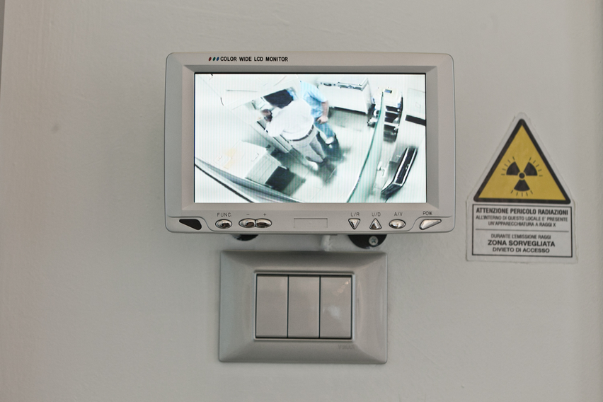
Who do the dentist and the staff leave the room when an x-ray is performed?
Although the dose of radiation for each single x-ray is extremely low, several x-rays a day over a long period of time would cause the dentist and the staff to be exposed to amounts of radiations well above the average.
If I am pregnant or I think I might be pregnant, should I postpone dental x-rays?
Do not forget to notify us if you are pregnant or if you think you might be pregnant. It is advisable to avoid x-rays during pregnancy, unless they are indispensable, because they harm the foetus even at low dosage and even when they do not involve the reproductive system.